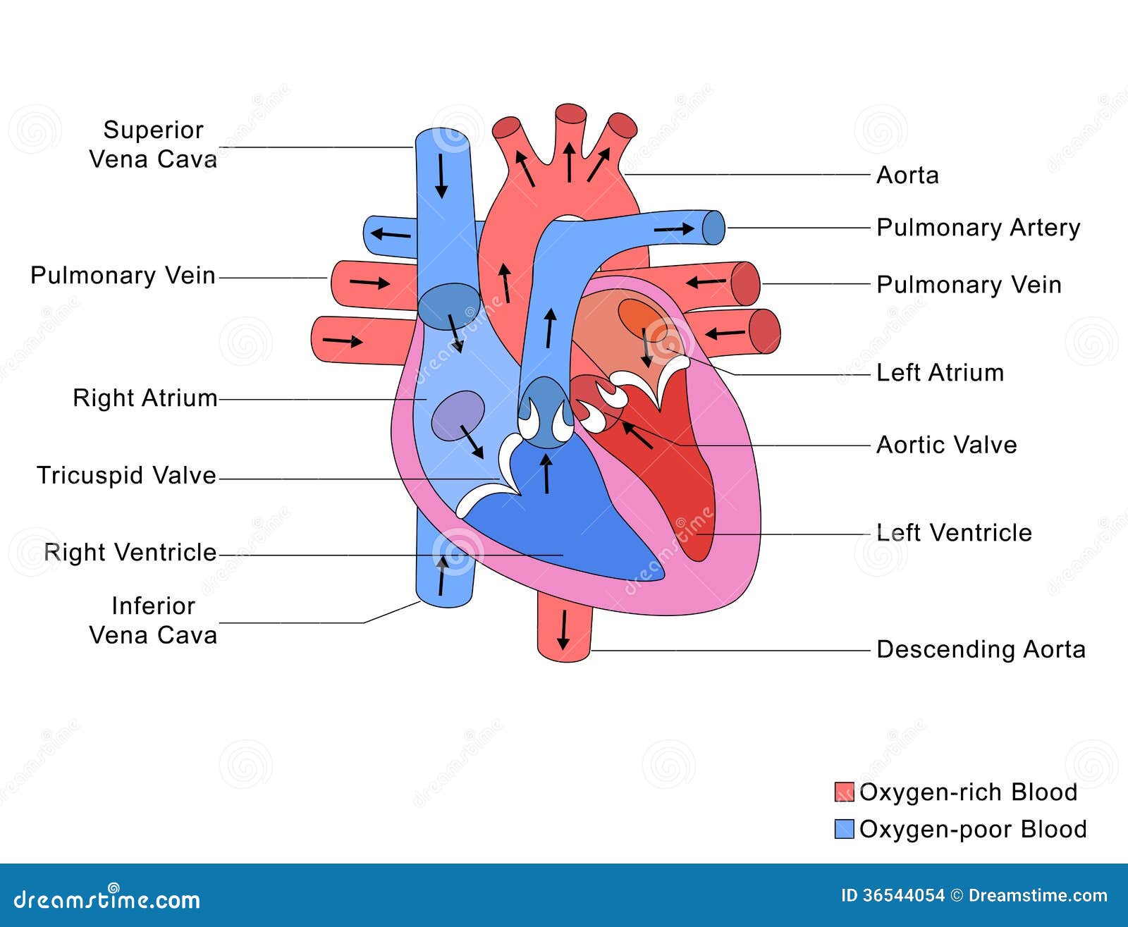
The two ventricles are thick-walled chambers that forcefully pump blood out of the heart. The two atria are thin-walled chambers that receive blood from the veins. The internal cavity of the heart is divided into four chambers: The outer layer of the heart wall is the epicardium, the middle layer is the myocardium, and the inner layer is the endocardium. Three layers of tissue form the heart wall. The visceral layer of the serous membrane forms the epicardium. The heart is enclosed in a pericardial sac that is lined with the parietal layers of a serous membrane. The left ventricle is thicker than the right ventricle as it takes more force to push blood to the body compared to the lungs.The human heart is a four-chambered muscular organ, shaped and sized roughly like a man's closed fist with two-thirds of the mass to the left of midline. The left atrium receives oxygenated blood from the lungs and the left ventricle pushes this oxygenated blood to the body. The right atrium receives deoxygenated blood from the body and the right ventricle pushes this deoxygenated blood to the lungs where it receives oxygen. The atria receive blood from veins and the ventricles push blood out of the heart into arteries. The heart itself needs its own blood supply for oxygen and nutrients as it is a muscle, so is supplied by the coronary arteries. 2 valves between the atria and the ventricles and 2 valves between the ventricles and the arteries that leave the heart.


The right and the left side of the heart are separated by the interventricular septum. In the heart there are 4 chambers the upper 2 chambers are called the atria and the bottom two chambers are called the ventricles.


 0 kommentar(er)
